


Cells 2023, 12(22), 2638; https://doi.org/10.3390/cells12222638 - 16 Nov 2023
Viewed by 1395
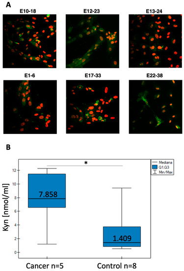
Abstract
►
Show Figures
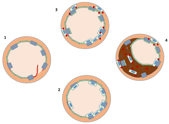
With the increasing demand for therapeutic antibodies, CHO cells have become the de facto standard as producer host cells for biopharmaceutical production. High production yields are required for antibody production, and developing a high-titer production system is increasingly crucial. This study was established
[...] Read more.
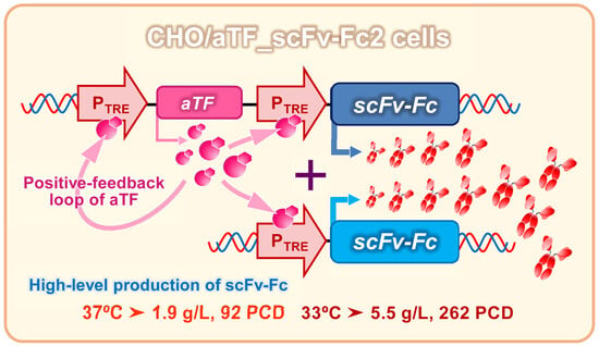
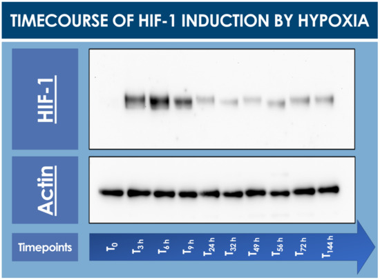
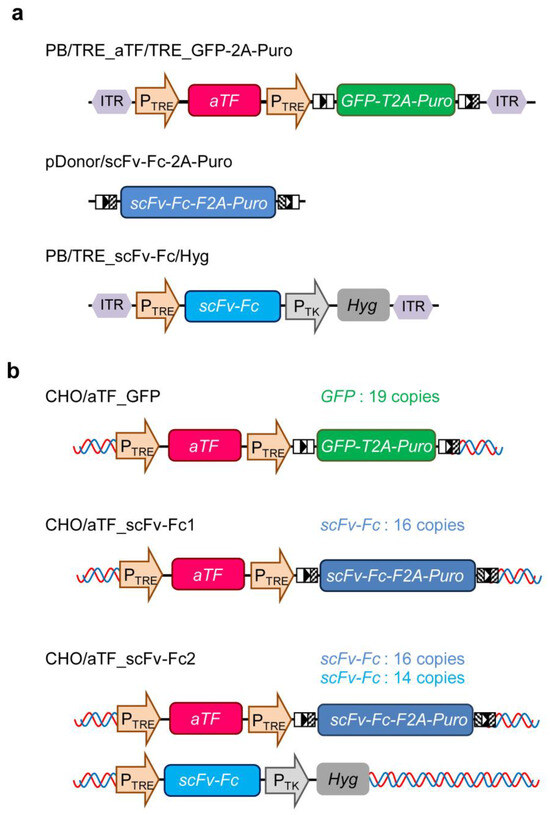
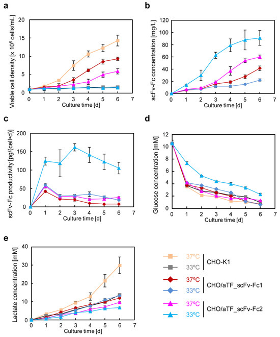
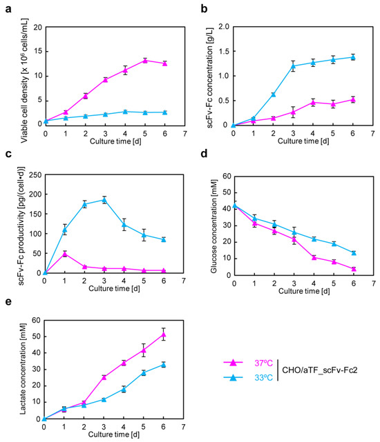
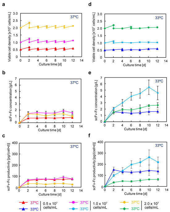
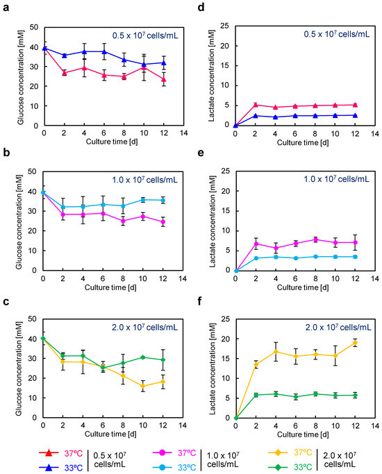
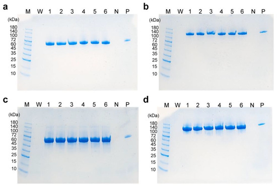
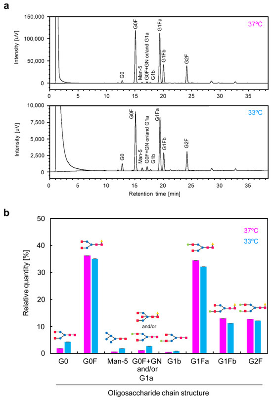
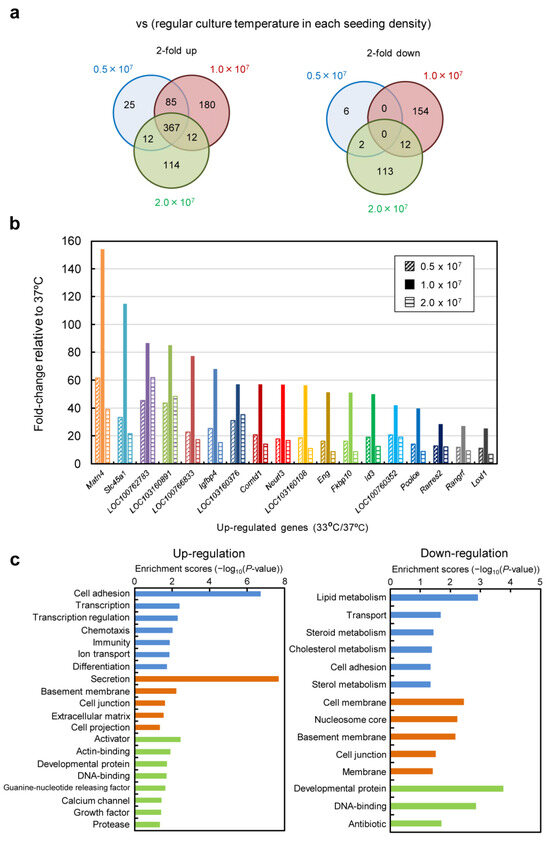
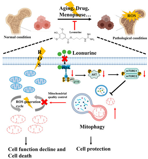
With the increasing demand for therapeutic antibodies, CHO cells have become the de facto standard as producer host cells for biopharmaceutical production. High production yields are required for antibody production, and developing a high-titer production system is increasingly crucial. This study was established to develop a high-production system using a synthetic biology approach by designing a gene expression system based on an artificial transcription factor that can strongly induce the high expression of target genes in CHO cells. To demonstrate the functionality of this artificial gene expression system and its ability to induce the high expression of target genes in CHO cells, a model antibody (scFv-Fc) was produced using this system. Excellent results were obtained with the plate scale, and when attempting continuous production in semi-continuous cultures using bioreactor tubes with high-cell-density suspension culture using a serum-free medium, high-titer antibody production at the gram-per-liter level was achieved. Shifting the culture temperature to a low temperature of 33 °C achieved scFv-Fc concentrations of up to 5.5 g/L with a specific production rate of 262 pg/(cell∙day). This artificial gene expression system should be a powerful tool for CHO cell engineering aimed at constructing high-yield production systems.
Full article
Graphical abstract
